
Cochlear implants and the Radiologist
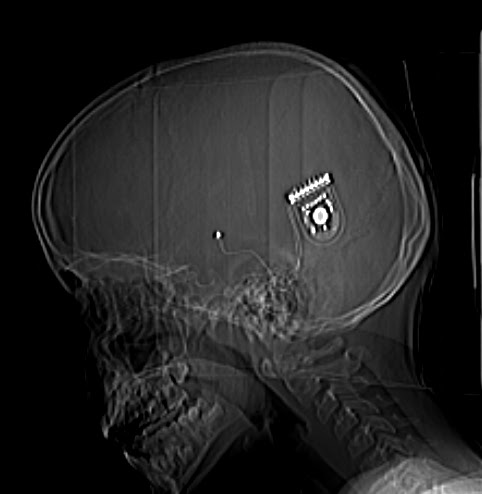
9 yr old girl shows presence of cochlear implant in situ, shows pneumocochlea, pneumocephalus, and subtle fluid density in to the middle ear location, possibly perilymph fistula, through the iatrogenic, intended cochleostomy. The electrodes appear close to cochlea particularly apical region and are normal. There is no evidence of infection on this. Treatment of such moderate air without parenchymal presence is usually conservative. Case and discussion by Dr MGK Murthy


Indication
Nonfunctional (ganglion cells ineffective)cochlea on both sides leading to bilateral sensorineural deafness with intact auditory nerve function. Usually in children, but of late the adult varieties are receiving the implant
Contraindications
· Obliterative labyrinthine ossification, severe cochlear or fenestrative otosclerosis, congenital cochlear malformations(mondini deformity, ossified cohlea etc), severe bilateral temporal bone fractures, Infected middle ear
· Usually HRCT temporal bone would evaluate all , but some prefer MRI to evaluate 8th nerve, speech and language functional MRI as well
Normal hearing
Sound from the environment—via the external and middle ear(including tympanic membrane and ossicles)--- Cochlea spiral ganglion cells----auditory nerve---brainstem.
Implant has two parts
External-microphone to pick the environment sound
-Speech processor-digitizes the signal
-Transmitter-converts to FM type of Radiosignals
Internal –kept aligned well by the magnets , and placed underskin behind ear
-Receiver/stimulator(disk shape)-converts FM signals to electric signals
- through a wire connected to Electrodes(usually 24)
-stimulate the spiral ganglion cells (apical better because sound will be more natural)------auditory nerve---brain. Sound produced is different from ordinary and robotic, needs adjustment, because 24 electrodes cannot match 15000 haircells normally present
Surgery
Making communication between mastoid/ middle ear and mastoid / cochlea . Intra -operative Radiographs help in correct positioning of the electrodes
Complications
Extra cochlear placement of the electrodes including in the semicircular canals, breakage facial palsy, infection, pneumocephalus, fluid drainage, meningitis, cochlear damage during insertion of electrodes, osteogenesis, vestibular symptoms etc
MR Safety
Previously thought to be Unsafe (because of the magnets with in the gadgets), recent models are classified as conditional (can be performed with specific recommendations of the manufacturer including the magnetic strength)
Cochlear implants and the Radiologist
 Reviewed by Sumer Sethi
on
Monday, June 27, 2011
Rating:
Reviewed by Sumer Sethi
on
Monday, June 27, 2011
Rating:
Reviewed by Sumer Sethi
on
Monday, June 27, 2011
Rating:
Sumer Sethi

Unique blend of academic excellence and entrepreneurship, heading leading firms in India- Teleradiology Providers, pioneering company providing teleradiology services and DAMS (Delhi Academy of Medical Sciences) Premier test preparation institute in India for MD/MS/MCI preparation. He has also been an invited faculty member at various conferences, including Teleradiology in IRIA 2008 and 2011, Hospital Build Middle East, Congress of the Brain Tumor Radiology in Neuro-oncology Society. Dr. Sethi is Editor-in-Chief of Internet Journal of Radiology. He has a keen interest in Web 2.0 technologies and in maintaining his famous radiology blog, which has been featured in multiple international journals.


No comments:
Post a Comment