
Iliotibial Band Syndrome-MRI
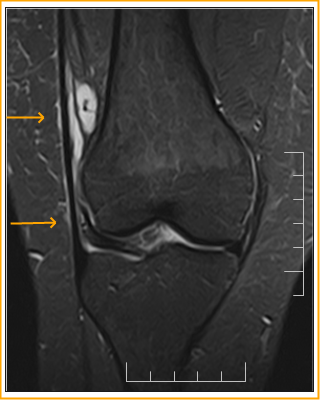
Patient presented with pain on the lateral aspect of knee, no injury.
Findings: Multilobulated cystic areas in the lateral recess deep to the iliotibial band along with focal discontinuity in the tract with communicating fluid space superficial to the band. Thickening of the tibial insertion of iliotibial band on the gerdy tubercle and there is reduced space between the distal iliotibial tract and lateral femoral condyle. These findings likely suggest iliotibial band friction syndrome with associated bursal cyst and possible discontinuity in the band.
Discussion : When the knee flexes, the ITB moves posteriorly along the lateral femoral epicondyle. When the band is excessively tight or stressed, the ITB rubs against the epicondyle irritating the lateral synovial recess.


Iliotibial Band Syndrome-MRI
 Reviewed by Sumer Sethi
on
Friday, September 11, 2015
Rating:
Reviewed by Sumer Sethi
on
Friday, September 11, 2015
Rating:
Reviewed by Sumer Sethi
on
Friday, September 11, 2015
Rating:
Sumer Sethi

Unique blend of academic excellence and entrepreneurship, heading leading firms in India- Teleradiology Providers, pioneering company providing teleradiology services and DAMS (Delhi Academy of Medical Sciences) Premier test preparation institute in India for MD/MS/MCI preparation. He has also been an invited faculty member at various conferences, including Teleradiology in IRIA 2008 and 2011, Hospital Build Middle East, Congress of the Brain Tumor Radiology in Neuro-oncology Society. Dr. Sethi is Editor-in-Chief of Internet Journal of Radiology. He has a keen interest in Web 2.0 technologies and in maintaining his famous radiology blog, which has been featured in multiple international journals.


No comments:
Post a Comment