
Leukodystrophy Teaching Points-MRI
One
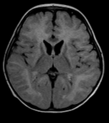
year male child born in caesaerian
delivery to non-consanguinous parents,
presents with abnormal movements and delayed milestones with no fever
are trauma. The MR imaging shows extensive centrifugal pattern of restricted
diffusion of myelin edema in supratentorial (posterior to the anterior
gradient) & infratentorial locations
with apparent “Tigroid” appearance with T2 shine through in nature with no
significant reduction in ADC with no mass effect or bleed or volume loss or circle of Willis flow
voids abnormality. Though the features are not specific, could suggest
leukodystrophies or metabolic etiologies. Case and teaching points by Dr MGK
Murthy.


Teaching
points :
- Various edema are vasogenic and (isotropically increased) or interstitial or cytotoxic and myelin (intramyelinic) varieties (isotropically restricted water diffusion).
- Absent or low grade myelin edema is seen in MPS, GM gangliosidoses, Zell Weger disease, Adrenomyeloneuropathy, L-2 hydroxyglutaric acid uria, non ketotic hyperglycinemia, classical phenyl ketonuria and Vander knap disease. Vanishing white matter or medium grade myelin edema is seen in metachromatic leukodystrophy (MLD), X-linked adrenoleukodystrophy and HMG co-enzyme lyase deficiency. High grade edema is seen in Krabbe disease, Canavan disease, hyperhomocystinemias andMaple syrup urine disease.
- MLD, globoid cell leukodystrophy , Canavan disease and X-linked ALD are genetically determined and referred to as classical varieties. Recent additions of leukodystrophies include megalencephalic leukoencephalopathy with subcortical cysts, Aicardi goutiere syndrome, vanishing white matter disease or leukodystrophy with brain stem and spinal cord involvement and high lactate.
- White matter diseases present with corticospinal tracts symptoms as against grey matter diseases (polio dystrophies) showing extrapyramidal symptoms.
- MLD variety shows progressive centrifugal white matter disease with posteroanterior gradiant with TIGROID pattern in centrum semiovale (due to sparing of the myelin around the transmedullary vessels). Krabbe disease suggests simultaneous and early involvement of supra and infratentorial regions.
Leukodystrophy Teaching Points-MRI
 Reviewed by Sumer Sethi
on
Tuesday, September 24, 2013
Rating:
Reviewed by Sumer Sethi
on
Tuesday, September 24, 2013
Rating:
Reviewed by Sumer Sethi
on
Tuesday, September 24, 2013
Rating:
Sumer Sethi

Unique blend of academic excellence and entrepreneurship, heading leading firms in India- Teleradiology Providers, pioneering company providing teleradiology services and DAMS (Delhi Academy of Medical Sciences) Premier test preparation institute in India for MD/MS/MCI preparation. He has also been an invited faculty member at various conferences, including Teleradiology in IRIA 2008 and 2011, Hospital Build Middle East, Congress of the Brain Tumor Radiology in Neuro-oncology Society. Dr. Sethi is Editor-in-Chief of Internet Journal of Radiology. He has a keen interest in Web 2.0 technologies and in maintaining his famous radiology blog, which has been featured in multiple international journals.


No comments:
Post a Comment