
Exostosis-not always easy to identify!
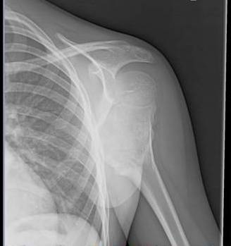
Young Skeleton with pain after fall shows exophytic opacity outwards from the metaphyseal location of humerus with undisplaced fractures in the vicinity with almost 2 Humeral heads appearance suggesting exostosis with suspicion for secondary chondrosarcoma of cartilage cap. MRI is needed for further evaluation.


Teaching points by Dr MGK Murthy, Dr Sumer Sethi.
· Defined as developmental dysplasia of peripheral growth plate which forms cartilage cap projection of bone near metaphysis of long bones. Peripheral chondroblast grows outwards acting as ectopic growth plate , stopping at maturity
· Most common benign bone tumor. Any bone forming in cartilage can get involved
· X ray hallmark is blending of tumor in to the underlying metaphysis along with calcification of cap elements
· Only 1% solitary ones can turn secondary chondrosarcoma at the cap , with 10% in multiple variety
· Cartilage cap measuring >1cm in adults and 2-3 cms in children on MRI along with sudden increase in bone scan uptake in adults is of concern
· Other complications include bursal formation
Exostosis-not always easy to identify!
 Reviewed by Sumer Sethi
on
Wednesday, December 07, 2011
Rating:
Reviewed by Sumer Sethi
on
Wednesday, December 07, 2011
Rating:
Reviewed by Sumer Sethi
on
Wednesday, December 07, 2011
Rating:
Sumer Sethi

Unique blend of academic excellence and entrepreneurship, heading leading firms in India- Teleradiology Providers, pioneering company providing teleradiology services and DAMS (Delhi Academy of Medical Sciences) Premier test preparation institute in India for MD/MS/MCI preparation. He has also been an invited faculty member at various conferences, including Teleradiology in IRIA 2008 and 2011, Hospital Build Middle East, Congress of the Brain Tumor Radiology in Neuro-oncology Society. Dr. Sethi is Editor-in-Chief of Internet Journal of Radiology. He has a keen interest in Web 2.0 technologies and in maintaining his famous radiology blog, which has been featured in multiple international journals.


1 comment:
It is also k/a OSTEOCHONDROMA.
It is not a TRUE benign tumor,
so
MOST COMMON TRUE benign tumor is Osteoid osteoma.
http://sumerdoc.blogspot.com/2011/05/osteoid-osteoma-ulna.html
Post a Comment