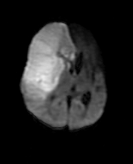
Hemimegalencephaly


Hemimegalencephaly or unilateral megalencephaly is a congenital disorder in which there is hamartomatous overgrowth of all or part of a cerebral hemisphere . The affected hemisphere may have focal or diffuse neuronal migration defects, with areas of polymicrogyria, pachygyria, and heterotopia. Hemimegalencephaly is a rare disorder and was first described by Sims in 1835.
Although the cause is unknown, it is postulated that it occurs due to insults during the second trimester of pregnancy, or as early as the 3rd week of gestation, as a genetically programmed developmental disorder related to cellular lineage and establishment of symmetry . Hemimegalencephaly may also be considered a primary disorder of proliferation wherein the neurons that are unable to form synaptic connections are not eliminated and are thus accumulated. No chromosomal abnormalities have been associated with hemimegalencephaly.
The isolated form, , occurs as a sporadic disorder without hemicorporal hypertrophy or cutaneous or systemic involvement.
The syndromic form is associated with other diseases and may occur as hemihypertrophy of part or all of the ipsilateral body. It has been described in patients with epidermal nevus syndrome, Proteus syndrome, neurofibromatosis type 1, hypermelanosis of Ito, Klippel-Weber-Trenaunay syndrome, and tuberous sclerosis. The third and least common type is total hemimegalencephaly, in which there is also enlargement of the ipsilateral half of the brainstem and cerebellum.
Epilepsy is the most frequent neurologic manifestation, occurring in greater than 90% of patients A characteristic finding is straightening of the ipsilateral frontal horn of the enlarged ventricle. At MR imaging, the white matter shows heterogeneous but frequently high signal intensity and there is often distinction of areas of agyria, pachygyria, and/or polymicrogyria. The white matter of the affected hemisphere may show advanced myelination for age . There is a roughly inverse relationship between the severity of the cortical and white matter abnormalities and the size of the cerebral hemisphere. Patients with agyria tend to have mild to moderate hemispheric enlargement, while those with polymicrogyria have more severe hemispheric enlargement .
Functional imaging with positron emission tomography has had good correlation with CT and MR imaging findings and has disclosed functionally abnormal brain regions in the noninvolved hemisphere that appeared structurally normal at CT and MR imaging.
Case by Dr MGK Murthy, Sr Consultant Radiologist
&
Dr.Sumer K Sethi, MD
Dr.Sumer K Sethi, MD
Consultant Radiologist ,VIMHANS and CEO-Teleradiology Providers
Editor-in-chief, The Internet Journal of Radiology
Director, DAMS (Delhi Academy of Medical Sciences
Hemimegalencephaly
 Reviewed by Sumer Sethi
on
Sunday, July 20, 2008
Rating:
 Reviewed by Sumer Sethi
on
Sunday, July 20, 2008
Rating:


No comments:
Post a Comment