
Congenital cytomegalovirus infection : CT Scan
History :
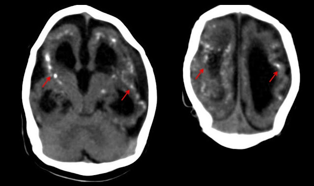
1 month neonate c/o convulsions
Findings:
CT brain shows ventricular dilatation
Periventricular calcification is seen around both the lateral ventricles
Diagnosis:
The findings are typical of congenital cytomegalovirus infection
Extra Edge Information
What is TORCH infection?
Toxoplasma, Rubella, Cytomegalovirus and herpes infection that typically affects newborn.
Types of calcification as clue to diagnosis?
CMV – Periventricular, Toxoplasma – basal ganglia
Other features of CMV infection?
Hepatosplenomegaly, chorioretinitis
 Reviewed by Sumer Sethi
on
Monday, February 14, 2022
Rating:
Reviewed by Sumer Sethi
on
Monday, February 14, 2022
Rating:
Unique blend of academic excellence and entrepreneurship, heading leading firms in India- Teleradiology Providers, pioneering company providing teleradiology services and DAMS (Delhi Academy of Medical Sciences) Premier test preparation institute in India for MD/MS/MCI preparation. He has also been an invited faculty member at various conferences, including Teleradiology in IRIA 2008 and 2011, Hospital Build Middle East, Congress of the Brain Tumor Radiology in Neuro-oncology Society. Dr. Sethi is Editor-in-Chief of Internet Journal of Radiology. He has a keen interest in Web 2.0 technologies and in maintaining his famous radiology blog, which has been featured in multiple international journals.


No comments:
Post a Comment