Pneumocystis Carnii-CT
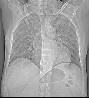
Clinical Profile : HIV positive status. There is presence of ground glass haziness with interstitial interlobular and intralobular septal thickening noted in both lungs with symmetrical and perihilar distribution, sparing the lung peripheries. No evidence any cystic lucencies are noted. Possibility of Pneumocystis Carnii. Case Submitted by Dr Swati Shah, MD, FRCR & Dr Sumer Sethi.
Features of P. Carnii on CT include:
- ground-glass pattern
- predominantly involving perihilar or mid zones
- there may be a mid, upper or lower zone predilection depending on whether the patient is on prophylactic aerosolised medication , if they are, then the poorly ventilated upper zones are prone to infection , whereas in those who are not the lower zones are more frequently involved
- reticular opacities or septal thickening may also be present
- a crazy paving pattern may therefore be seen when both ground-glass opacies and septal thickening are superimposed on one another
- pneumatocoeles
- pleural effusions are rare
Pneumocystis Carnii-CT
 Reviewed by Sumer Sethi
on
Tuesday, June 26, 2012
Rating:
 Reviewed by Sumer Sethi
on
Tuesday, June 26, 2012
Rating:


1 comment:
Dear sir,
Nice case....
but how will we differentiate it from the evolving Pulm odema. on plain chest xray: if the b/l symm. middle zones getting involved...n sparing the lung peripheries n heart size normal..
thanks in advance...
Post a Comment