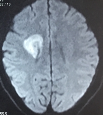
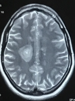
Case Report: 23 year old left hemiparesis. Clinical diagnosis MS. CSF OCB positive . Poor response to steroid. CEMR images are provided.MRI images reveal rounded lesion with alternating layers of increased and reduced signal, along with diffusion restriction. Case Submitted by Dr Rahul Rajeev, DM (Neurology std)
Quick Notes: Balo concentric sclerosis is a rare and severe monophasic demyelinating disease, considered a subtype of multiple sclerosis, appearing as a rounded lesion with alternating layers of increased and reduced signal giving it a characteristic 'bulls eye or 'onion bulb' appearance
 |
| FLAIR AXIAL MRI |
 |
| DW-MRI AXIAL |
 |
| CEMR |
 |
| AXIAL T2 WI |


 Reviewed by Sumer Sethi
on
Thursday, March 29, 2018
Rating:
 Reviewed by Sumer Sethi
on
Thursday, March 29, 2018
Rating:


No comments:
Post a Comment